The anatomy of the knee
The knee is a joint that makes flexion and extension movements possible. The three bone segments that make up the joint are: the femur, tibia and patella. In its distal portion the femur is composed of two different structures, the condyles (medial and lateral), with a convex surface, which are free to slide and roll on the two tibial hemiplateaus.
The surface of the tibia, called the tibial plateau, is also made up of two symmetrical parts , the tibial hemiplateaus (medial and lateral), with a concave surface. The flexion-extension mechanism is the result of the rolling and sliding of the convex surface of the femur over the surface of the concave tibial hemiplateaus.
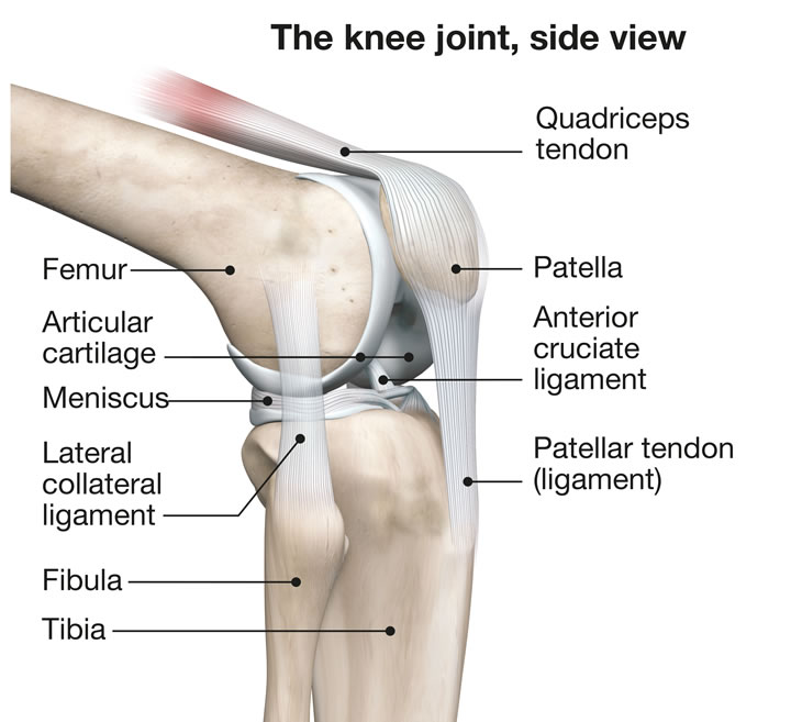
The patella is located in the anterior region of the knee. It has an anterior surface and a posterior surface that slides, during flexion and extension, within the femoral intercondylar groove, a groove on the anterior side of the femur, between the femoral and medial condyles. The patella plays a fundamental role in the extensor system of the knee, since the quadriceps muscle is inserted at its superior pole and the patellar tendon originates from its inferior pole.
The joint as a whole is covered by the joint capsule, a sturdy membrane that contains synovial fluid. In addition to the muscles that strengthen the joint and allow movement, the “scaffolding” of the knee is made up of ligaments, which withstand stress forces and stabilize it.
The cruciate ligaments are located in the middle of the knee and stabilize the femur and tibia in an anterior-posterior direction.
The collateral ligaments are located on either side of the knee and stabilize the femur and tibia against varus and valgus stresses.
The function of the menisci is very important in the structure of the knee. These are two fibrocartilages that are interposed between the two femoral condyles and the two tibial hemiplateaus. The medial and lateral menisci, shaped like an internally facing C, have a triangular cross-section and increase the congruence between the articular surfaces of the femur and tibia, acting as protective “pads” for the cartilage.
The information provided is not medical advice, nor is it intended as a substitute for medical advice. Under no circumstances should this information be a substitute for a consultation, examination or diagnosis given by a doctor.
2024 Gruppo Bioimpianti s.r.l. . All right reserved. Made with by SYROOP.
GRUPPO BIOIMPIANTI s.r.l. – Via Liguria 28, 20068 Peschiera Borromeo (Milan) – Italy
P.IVA: 10617240154, DUNS 443386602


